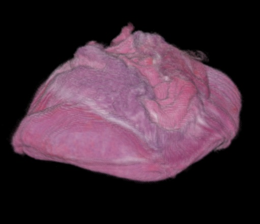
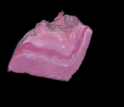
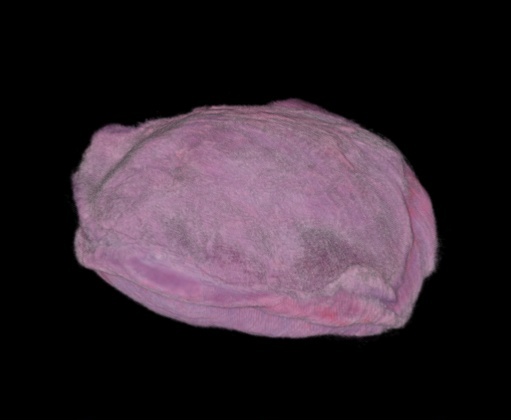
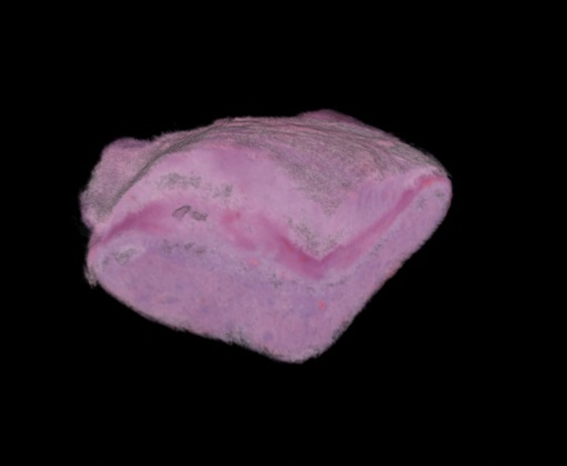
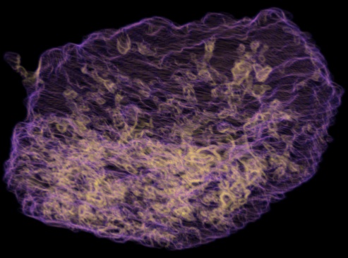
Below, you're looking at digital mouse placenta, originally coming from something like this (the blob on the bottom).
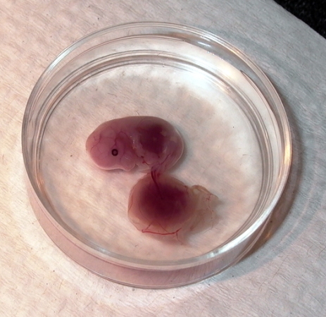
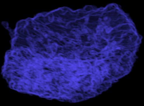
Biologists will take the placenta, mount it in paraffin, and slice it up, and scan in a bunch of .png files. You usually end up with about 1 TB of images per placenta. What you see below are two digital reconstructions of the placenta that can be diced and sliced digitally.
| Full Placenta | Cropped |
|---|---|
|
|
|
|
These images show the infiltrations and extrusions of the labyrinth interface. In general, the more brightly colored the features, the more they stick in or stick out.
| 05_1903 -- Wildtype | 05_1904 -- Mutant | |
|---|---|---|
| Full Labyrinth | 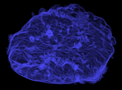 |
 |
| Infiltrations | 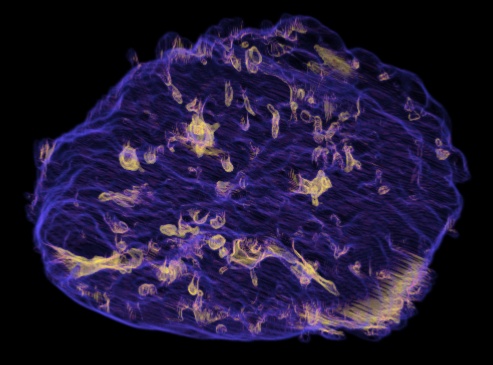 |
 |
| Extrusions | 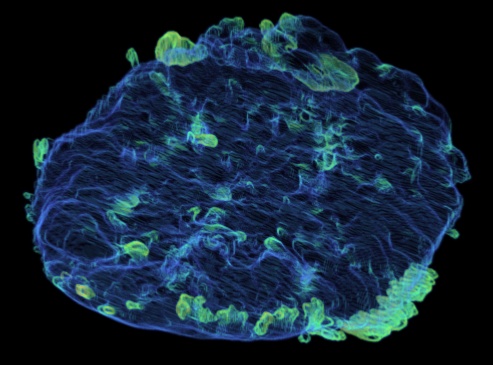 |
 |
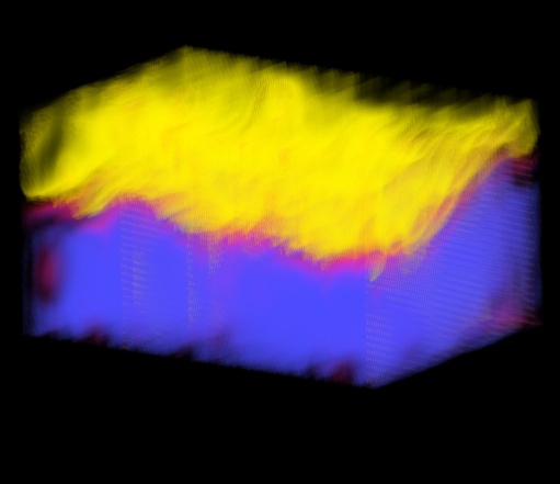
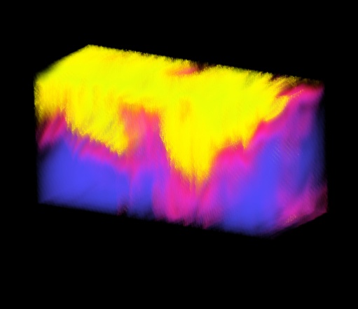
The images below show the labyrinth (blue), spongiotrophoblast (yellow) and the boundary (magenta) between them.
| Wildtype (thin interface) | Mutant (thick interface) |
|---|---|
 |
 |